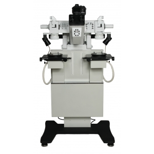
PELENG MC-4 Comparison Microscope
|
PURPOSE
|
|
|
|
|
SOFTWARE
|
|
|
|
|
OPERATION MODES
|
|
|
|
|
SPECIFICATIONS
|
|
| Magnification for direct-view optics, power, with max error of 5% | 5; 10; 20; 40; 80 |
| Field of view, mm, min | 40; 20; 10; 6; 2,5 |
| Integrated USB 2.0 colour TV camera, MP | 5,0 |
| Digital photo camera, MP | 12,0 |
| Focussing range, mm, min | 15 |
| Objective to object distance, mm | 115 ± 10 |
| Eyepiece diopter adjustment range, dpt | +5...-5 |
| Pupil distance adjustment range, mm | 56+1,0...75-1,0 |
| Sleeves and bullets diameter, mm | 3 ÷ 25 |
| Sleeves and bullets length, mm | 4 ÷ 70 |
| Distance and element measurement range, mm | 0 ÷ 60 |
| Scale division value, mm | 0,05 |
| Linear dimension measurement error, mm | 0,05 |
| Power supply voltage (50 Hz), V | 230 ± 23 |
| Power consumption, W, max | 650 |
| Motorized stage movement speed, mm/s , max | 5 |
| Eypiece height (from floor), mm | 1160 |
| Overall dimensions, mm | 680×600×1210 |
| Weight, кg | 130 |
|
Stage movement:
|
|
| - in XY plane from central position, mm | ± 20 |
| - Z axis rotation, º | 360 |
| - along Z axis, mm | 200 ± 10 |
| - motorized shift of both stages along Y axis (left-right from central position), mm | 50 ± 5 |
